
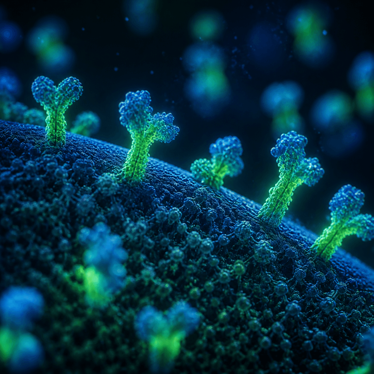
Laboratories identify mesenchymal stem cells by the proteins on the outside of the cell membrane. CD105, CD73, and CD90 form the positive side of the standard panel, and a second list of markers has to be nearly absent. This guide covers what surface marker testing measures and where its limits sit.
TLDR: The International Society for Cellular Therapy set minimum criteria for mesenchymal stem cells in 2006. At least 95 percent of the population must express CD105, CD73, and CD90, and no more than 2 percent may express CD45, CD34, and several other markers. Flow cytometry produces those percentages. The test confirms identity and purity in one sample at one moment. It does not measure potency, and banking adipose tissue does not guarantee eligibility, access, or benefit later, since these cells are investigational and not FDA approved.
Important Disclaimer: Save My Fat is a tissue preservation and connection service. It does not provide FDA-approved treatments or cures, and it does not guarantee eligibility, access, or clinical benefit from any present or future therapy. The marker testing described here is laboratory characterization, not a clinical service and not a predictor of any result. This article is for educational purposes only. Readers should consult their own licensed healthcare professionals before making decisions about tissue banking.
A vial of frozen cells looks like every other vial of frozen cells. Even under a microscope, one stromal cell looks much like the next. So laboratories do not identify these cells by appearance. They identify them by the proteins studding the cell surface, then count how many cells carry each one.
That counting exercise is the backbone of cell identity testing, and it explains why three marker names appear on nearly every characterization report in this field. As of July 2026, one state has written those reports into statute. This guide covers the panel, the instrument behind it, and the limits.
What CD105, CD73, and CD90 Say About Mesenchymal Stem Cells
In 2006, a committee at the International Society for Cellular Therapy published a short paper so laboratories would stop applying one name to very different cells. Dominici and colleagues set three minimum criteria in the ISCT minimal criteria statement: plastic adherence in culture, a defined surface marker profile, and differentiation to osteoblasts, adipocytes, and chondroblasts.
The marker half of that definition carries numbers. At least 95 percent of the population must express CD105, CD73, and CD90. No more than 2 percent may express CD45, CD34, CD14 or CD11b, CD79alpha or CD19, and HLA-DR. Flow cytometry measures both figures, which is how mesenchymal stromal cells get told apart.
| Marker | Panel side | ISCT threshold |
|---|---|---|
| CD105 | Positive | 95 percent or more |
| CD73 | Positive | 95 percent or more |
| CD90 | Positive | 95 percent or more |
| CD45, CD34 | Negative | 2 percent or fewer |
| CD14 or CD11b, CD79alpha or CD19, HLA-DR | Negative | 2 percent or fewer |
Read that table as a pass or fail gate, not a score. A sample at 91 percent for CD90 does not partially qualify, and neither does one at 6 percent for CD45. The panel is also only one of the three criteria.
How Flow Cytometry Works, in Plain Language
Flow cytometry sounds forbidding, but the idea underneath is counting. A technician suspends the sample in fluid and adds antibodies. Each antibody is built to stick to one specific surface protein, and each carries a fluorescent dye that glows a particular color when the right wavelength of light strikes it.
The instrument pushes that fluid through a channel narrow enough that cells line up and pass one at a time, single file. A laser hits each cell in turn. If antibodies are bound to it, the dye lights up, and detectors around the stream record color and brightness. Unlabeled cells pass through dark.
Software tallies the run. Out of tens of thousands of cells counted, it reports the percentage that lit up for each marker, and those percentages are what the 95 percent and 2 percent thresholds describe. The instrument is not grading quality. It is counting which proteins the cells carry.
Why the Negative Markers Matter as Much as the Positive Ones
Most patient-facing writing stops at the positive three, which misses half the point. CD45 sits on cells of the blood-forming lineage. CD34 appears on hematopoietic progenitors and on cells lining blood vessels. CD14 and CD11b mark monocytes and macrophages, CD79alpha and CD19 mark B cells, and HLA-DR marks antigen-presenting immune cells.
That matters because adipose tissue is dense with blood vessels and resident immune cells. A sample with strong CD105, CD73, and CD90 signal but 20 percent CD45 is not a clean stromal population. It is stroma mixed with blood-lineage cells. The negative markers are the purity check, and a report omitting them is incomplete.
What Surface Marker Testing Does Not Tell You
Here is the honest boundary. Surface marker testing is an identity and purity check on one sample, drawn at one moment, under one set of laboratory conditions. It answers what a cell population appears to be. It does not answer what that population can do, and it never has.
| Answers | Does not answer |
|---|---|
| Share of the sample carrying CD105, CD73, CD90 | Whether the cells would benefit a patient |
| How much blood-lineage contamination is present | How potent the sample is |
| Whether it matches an accepted identity definition | Whether an approved therapy will exist |
| The condition of this sample today | How it behaves years from now |
Every entry on the right stays open no matter how clean the report looks. The FDA’s consumer alert on regenerative medicine products states that blood-forming stem cells from cord blood are the only approved stem cell products, and that these products are not approved for arthritis or Alzheimer’s.
Where Surface Marker Reports Became a Legal Requirement
For twenty years, marker testing was a scientific convention rather than a legal duty. That shifted in 2026. Tennessee Public Chapter 1016, signed May 19, 2026 and effective July 1, 2026, sets product standards for regenerative medicine therapy: the manufacturer must validate its isolation techniques with cell viability and surface marker reports, per the Tennessee text.
Arizona nearly followed. SB 1214 would have imposed comparable manufacturer validation duties, including viability and surface marker reports, but Governor Katie Hobbs vetoed the bill on June 19, 2026 and no override followed. So one state requires the report. Everywhere else, asking for it is diligence rather than compliance.
What This Means for Banked Adipose Tissue
Two details keep this from mapping neatly onto banked tissue. The ISCT criteria describe cells grown in culture, so applying the 2 percent ceiling to fresh, uncultured stromal vascular fraction compares two different things. The joint IFATS and ISCT statement addresses that gap, anticipating colony-forming unit fibroblast frequency above 1 percent for that fraction and above 5 percent once expanded.
Markers are also not permanent. Wagner and colleagues found that within 43 to 77 days of cultivation, cells showed attenuated expression of specific surface markers alongside morphological changes. Devitt and colleagues examined samples stored from 2 to 1,159 days and reported that MSC markers were maintained throughout. Both are preliminary findings, not clinical evidence.
Practically, a marker report generated years from now on expanded cells is a different document from one generated on a fresh sample today. Knowing how adipose tissue biobanks document samples, and how cryopreservation works, gives that comparison context that a licensed physician should interpret.
Frequently Asked Questions
What do CD105, CD73, and CD90 markers actually indicate about a cell?
They indicate the cell carries the surface protein profile the field associates with mesenchymal stromal cells. Under the ISCT criteria, at least 95 percent of a population must express all three. That is a statement of identity, not of what the cells will do in a person.
Why must cells also test negative for markers like CD45 and CD34?
Because the positive panel alone cannot detect contamination. CD45 marks blood-forming lineage cells, and CD34 appears on hematopoietic progenitors and vessel-lining cells. Without the 2 percent ceiling on the negative markers, a sample could post strong CD105, CD73, and CD90 numbers while still being substantially blood and endothelial cells.
How is this marker testing performed in a laboratory?
By flow cytometry. Cells are suspended in fluid with antibodies that each bind one surface protein and carry a fluorescent dye. The instrument moves the fluid through a narrow channel so cells pass a laser single file. Detectors record which cells fluoresce, and software reports the percentages.
Does surface marker testing happen during Save My Fat’s banking process?
Save My Fat does not operate a laboratory and does not collect, process, store, or test tissue. Processing and storage are handled by a U.S.-based tissue bank. Which characterization assays run on a sample depends on the laboratory’s protocols and the intended use, and Save My Fat does not interpret those tests.
Does having these markers mean a cell will provide clinical benefit?
No. Marker expression is an identity and purity measurement, not a potency or efficacy measurement. Adipose-derived cells remain investigational and are not FDA approved for any condition, the available evidence is preliminary, and outcomes cannot be predicted. Decisions belong with a licensed physician who knows the patient.
Do surface markers stay the same during storage and culture?
Not necessarily. Extended culture has been associated with attenuated expression of specific surface markers alongside morphological changes. Storage appears more stable in published characterization work, with MSC markers maintained across a wide range of durations. Both are laboratory observations, and more research is needed.
Key Takeaways
Surface marker testing exists because these cells cannot be told apart by eye. The ISCT criteria published in 2006 set a workable definition: at least 95 percent of a population expressing CD105, CD73, and CD90, no more than 2 percent expressing CD45, CD34, and the rest of the negative panel, plus plastic adherence and trilineage differentiation.
Flow cytometry supplies those percentages by counting cells one at a time past a laser. The negative markers do as much work as the positive ones, because they are the part of the panel that detects blood-lineage and vascular contamination. What the test cannot do is measure potency or predict benefit. Tennessee’s 2026 statute made manufacturer surface marker reports a legal requirement there, though it remains one state.
Save My Fat connects patients and providers with a U.S.-based tissue bank. It is not a tissue bank, a laboratory, a medical practice, or a treatment provider. Stem cell and regenerative medicine regulations vary by state, including specific informed-consent and disclosure requirements in Florida, Utah, California, Georgia, and Tennessee governing tissue and stem cell services. Banking adipose tissue does not connect patients to any treatment pathway, and any future use depends on FDA regulatory status, physician guidance, and the availability of approved or investigational pathways at that time.
Patients weighing whether to bank adipose tissue can review current plans or reach the team through the contact page with questions about documentation and storage.
Save My Fat works with a U.S.-based tissue bank for laboratory processing and storage.
This article is for educational purposes only and does not constitute medical or legal advice. Please consult your own licensed healthcare provider before making any decisions about banking, treatment, or research participation.
About the author: Oscar Tellez is the founder and CEO of Save My Fat. He holds a Bachelor of Science in Exercise Science and Health Promotion from Florida Atlantic University. He has spent a decade in the regenerative medicine industry across product distribution, laboratory and vendor relationships, and provider training. He is not a licensed clinician, and this article is educational, not medical advice.
Related guide: the stem cell science glossary.