Trilineage differentiation in mesenchymal stem cells is the functional test that separates a real stem cell population from ordinary connective tissue cells that look identical in a flask. It asks whether one population can become bone, fat, and cartilage cells in a dish. This guide covers the three assays, the stains and biomarkers behind each, and the hard limit on what a passing result means.
TLDR: Trilineage differentiation is the third of the three ISCT minimal criteria published by Dominici and colleagues in 2006, alongside plastic adherence and a surface marker profile. A qualifying population must become osteoblasts, adipocytes, and chondroblasts in vitro. Each lineage carries its own stain and biomarker panel: alizarin red or von Kossa for bone, oil red O or Nile red for fat, alcian blue or safranin O for cartilage. This is an identity and quality test run in a dish, not evidence of clinical benefit, and banking adipose tissue does not guarantee eligibility, access, or clinical benefit.
Important Disclaimer: Save My Fat is a connector service linking patients and providers to a U.S. tissue bank. Save My Fat does not provide FDA-approved treatments or cures and does not guarantee eligibility, access, or clinical benefit. Trilineage differentiation is a laboratory characterization method, and adipose tissue and stromal vascular fraction remain investigational and not FDA approved. This content is for educational purposes only, and readers should consult their own licensed healthcare professionals.
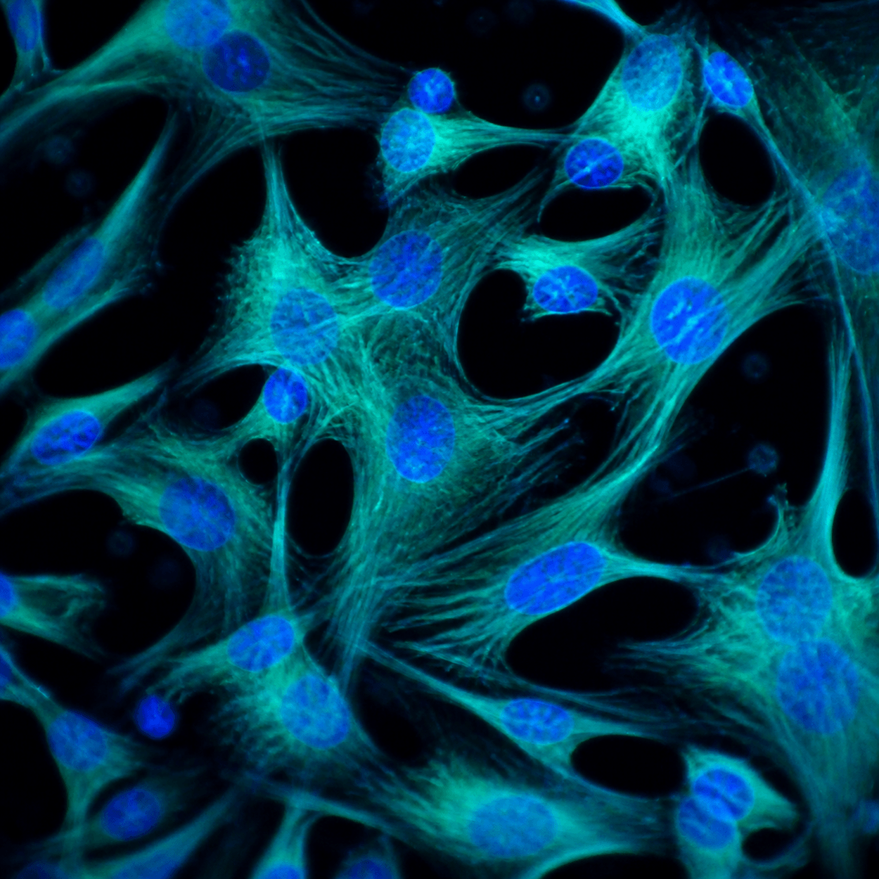
A mesenchymal stem cell and a skin fibroblast look nearly identical in a culture flask. Both are spindle shaped, both stick to plastic, and both keep dividing. Surface markers settle less than people assume, since unrelated cell types carry many of the same ones.
So the field stopped asking what a cell looks like and started asking what it can do. That produced the third and hardest ISCT criterion, and it explains why the phrase “stem cell” on a marketing page means little without a named assay behind it.
What Is Trilineage Differentiation in Mesenchymal Stem Cells
Differentiation is a general purpose cell becoming a specialized one, and a mesenchymal stromal cell in a flask is uncommitted. One chemical recipe moves it toward bone, a second makes it fill with lipid droplets, and a third, under specific physical conditions, starts it laying down cartilage matrix.
Trilineage means all three directions tested in parallel from one starting population. The laboratory splits the culture three ways and applies a different induction medium to each arm. In vitro means the work happens outside a body, under fully controlled conditions. New readers can start with what mesenchymal stromal cells are.
The Third of Three Mesenchymal Stem Cell Identification Criteria
The standard comes from Dominici and colleagues, published in Cytotherapy in 2006, and it states all three requirements in a single passage: “First, MSC must be plastic-adherent when maintained in standard culture conditions. Second, MSC must express CD105, CD73 and CD90, and lack expression of CD45, CD34, CD14 or CD11b, CD79alpha or CD19 and HLA-DR surface molecules. Third, MSC must differentiate to osteoblasts, adipocytes and chondroblasts in vitro.”
The first two describe a cell at rest. Flow cytometry sets the thresholds at 95 percent or more for CD105, CD73, and CD90, and 2 percent or less for the excluded markers. The third asks the cell to perform. The ISCT minimal criteria are a floor for identification, never a statement about what the cells do inside a person.
The Three Assays, Their Stains, and Their Biomarkers
The joint statement from the International Federation for Adipose Therapeutics and Science and the ISCT, published by Bourin and colleagues in 2013, pairs a histology stain with a biomarker panel for each lineage. A credible differentiation claim rests on both. The table below is that checklist.
| Lineage | Histology stain | Biomarkers reported |
|---|---|---|
| Osteogenic, bone forming | Alizarin red or von Kossa | Alkaline phosphatase, bone sialoprotein, osteocalcin, osterix, runx2 |
| Adipogenic, fat storing | Oil red O or Nile red | Adiponectin, C/EBP alpha, FABP4, leptin, PPAR gamma |
| Chondrogenic, cartilage forming | Alcian blue or safranin O | Aggrecan, collagen type II, Sox 9 |
Two stains appear per lineage because either one is accepted, not because both are required. The biomarker column matters as much as the color.
Osteogenic: alizarin red or von Kossa
Osteogenic induction pushes cells toward osteoblasts, which lay down bone matrix. Alizarin red binds calcium deposits and turns them deep red, while von Kossa targets phosphate and renders deposits black or brown. Runx2 and osterix switch the program on.
Adipogenic: oil red O or Nile red
Adipogenesis is the easiest of the three to see, as cells stop looking spindly, round up, and fill with lipid droplets. Oil red O stains those droplets bright red, and Nile red is a fluorescent alternative for quantitative work. PPAR gamma and C/EBP alpha drive the program.
Chondrogenic: alcian blue or safranin O, in pellet or micromass culture
Chondrogenesis is the hardest of the three. These cells will not do it spread flat across plastic. They have to be crowded, so the assay runs in pellet or micromass culture, a dense clump mimicking developing cartilage. Alcian blue binds glycosaminoglycans and safranin O stains proteoglycans.
CFU-F Frequency, the Number Behind the Population
A stain shows that some cells in a well went where they were pushed, not how many cells in the starting material were capable of going anywhere. The colony forming unit fibroblast assay answers that, by plating cells at low density and counting colonies from single founders.
The joint statement sets the expectation plainly. Anticipated CFU-F frequency is greater than 1 percent for stromal vascular fraction and greater than 5 percent for culture expanded adipose-derived stromal cells. The IFATS and ISCT joint statement treats those as quality control figures, not performance claims. Mitchell and colleagues reported colony-forming unit fibroblasts in initial SVF “at a frequency of 1:32”, roughly 3.1 percent, in Stem Cells.
What a Passing Result Does Not Prove
Here is the sentence marketing copy leaves out. Trilineage differentiation is a laboratory identity and quality test. It tells you what a population of cells is, not what those cells do inside a person, and it is not evidence of clinical benefit.
The clinical record shows it. In the ADIPOA2 trial, culture expanded autologous adipose-derived MSCs were injected once into knees with mild to moderate osteoarthritis, against placebo. At six months, 47.3 percent of the cell group and 54.8 percent of the placebo group met the strict OARSI/OMERACT responder definition, a relative risk of 0.86 with a P value of.46. Pers and colleagues concluded that the injection “did not significantly improve pain and function”. Those cells met the laboratory definition, and the trial was still negative.
The FDA’s consumer alert on regenerative medicine products names arthritis, back pain, Parkinson’s disease, ALS, Alzheimer’s disease, cardiovascular and lung disease, autism, and blindness among conditions these products are not approved for. Adipose-derived cells are under clinical investigation, the evidence is preliminary, and a licensed provider is the right person to interpret it.
Why “Stem Cell” Means Nothing Without the Assay Behind It
A trilineage result is a snapshot of one batch at one moment. Bonab and colleagues reported that “differentiation potential dropped from the 6th passage on”, with average population doublings falling from 7.7 to 1.2 by the tenth passage, in BMC Cell Biology. A differentiation report without a passage number is half a result.
So the useful question is never whether a company says stem cells. It is which criteria were measured, on which batch, at which passage, and at what CFU-F frequency. Readers weighing tissue types can review how MSC sources compare, while storage turns on separate measurements described under cryopreservation and tissue measurement.
Frequently Asked Questions
What does trilineage differentiation actually mean in plain language?
It means one population of cells can be pushed down three specialized paths in a laboratory dish. Given the right chemical recipe, those cells become bone-forming osteoblasts, fat-storing adipocytes, or cartilage-forming chondroblasts. All three arms run in parallel from one starting population.
Why do scientists use this test to confirm a cell is a true mesenchymal stem cell?
Because surface markers alone are not enough. Fibroblasts and other connective tissue cells share much of the same profile and look identical in a flask. Differentiation is a functional demand rather than a description, so a population meeting it has shown the defining capability.
How does this relate to the ISCT minimal criteria?
It is the third of the three. The first is plastic adherence in standard culture conditions. The second is a surface marker profile measured by flow cytometry, at 95 percent or more for CD105, CD73, and CD90 and 2 percent or less for the excluded markers.
What is CFU-F frequency and why does it appear alongside the stains?
The colony forming unit fibroblast assay counts how many cells in a preparation can start a colony on their own. The IFATS and ISCT joint statement anticipates greater than 1 percent for stromal vascular fraction and greater than 5 percent for culture expanded cells. It describes the preparation only.
What would happen if a cell failed the trilineage differentiation test?
The population would not meet the ISCT definition of a mesenchymal stromal cell, whatever its markers looked like. A failure usually points to something identifiable, such as a mixed population, too many passages, or an induction protocol that did not work.
Does Save My Fat perform differentiation testing on banked tissue?
No. Save My Fat is a connector linking patients and providers to a U.S. tissue bank, and it does not collect, process, store, or treat tissue. Trilineage assays are research methods requiring cells isolated and expanded in culture first. Outcomes cannot be predicted.
Key Takeaways
Trilineage differentiation is the third ISCT minimal criterion and the only one asking a cell to perform rather than be counted. A qualifying population must become osteoblasts, adipocytes, and chondroblasts in vitro. Bone is documented with alizarin red or von Kossa, fat with oil red O or Nile red, and cartilage with alcian blue or safranin O in pellet or micromass culture, each paired with its own biomarker panel.
CFU-F frequency completes the picture, anticipated above 1 percent for stromal vascular fraction and above 5 percent for culture expanded cells. Capacity drifts with passage number, so a report without one is incomplete. The limit matters most: this is an identity and quality test in a dish, not evidence of clinical benefit. Adipose-derived cells remain investigational and not FDA approved, and outcomes cannot be predicted.
Save My Fat connects patients and providers with a U.S.-based tissue bank. It is not a tissue bank, a laboratory, a medical practice, or a treatment provider. Stem cell and regenerative medicine regulations vary by state, including specific informed-consent and disclosure requirements in Florida, Utah, California, Georgia, and Tennessee governing tissue and stem cell services. Banking adipose tissue does not connect patients to any treatment pathway, and any future use depends on FDA regulatory status, physician guidance, and the availability of approved or investigational pathways at that time.
Anyone weighing adipose tissue banking for potential future use can review current pricing or contact our team with questions about how collection works.
Save My Fat works with a U.S.-based tissue bank for laboratory processing and storage.
This article is for educational purposes only and does not constitute medical or legal advice. Please consult your own licensed healthcare provider before making any decisions about banking, treatment, or research participation.
About the author: Oscar Tellez is the founder and CEO of Save My Fat. He holds a Bachelor of Science in Exercise Science and Health Promotion from Florida Atlantic University. He has spent a decade in the regenerative medicine industry across product distribution, laboratory and vendor relationships, and provider training. He is not a licensed clinician, and this article is educational, not medical advice.
Related guide: the stem cell science glossary.